Diagram Of Hip.and Back.muscles : Iliopsoas Wikipedia
Diagram Of Hip.and Back.muscles : Iliopsoas Wikipedia. The part of the nerve that emerges out of the spine is called the nerve root. If you are starting to feel hip pain or stiffness, you'll want to know more about the bones and muscles that make up the hip's anatomy. The four groups are the anterior group, the posterior group, adductor group, and finally the abductor group. The hip abductors consist of the: Female hip muscles diagram ~ anatomy of the male and female pelvis comprehensive orthopaedics.
ads/bitcoin1.txt
Five pairs of lumbar spinal nerves labeled l1 to l5 branch off your spinal cord and exit through small holes between the vertebrae. Related posts of muscles of the lower back and hip diagram muscle anatomy chart. The hip joint is made up of two. Muscles of the gluteal region. The main nerves of the hip that supply the muscles in the hip include the femoral, obturator, and sciatic nerves.
How Underactive Gluteal Muscles Can Cause Lower Back Pain Lifemark from www.lifemark.ca It runs from your lower back through your pelvis, passing to the front of your hip where it attaches to the top of your femur, which is your thigh bone. To learn more about the anatomy of the spine, watch this video. Related posts of muscles of the lower back and hip diagram muscle anatomy chart. The psoas muscle is a deep muscle that connects your spine to your leg.in fact, it's the only muscle that does so. The bones of the hip include the femur, the ilium, the ischium, and the pubis. The thigh bone or femur and the pelvis join to form the hip joint. The hip abductors consist of the: Common causes of tight hip and lower back muscles include injury, too little activity, too much activity and muscular imbalances.
As you can see from the diagram to the right, there are many muscles and tendons that make up the hip and buttocks region.
ads/bitcoin2.txt
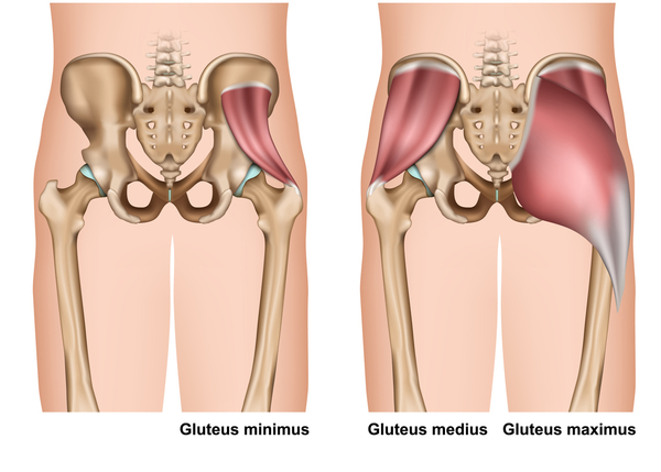
The four groups are the anterior group, the posterior group, adductor group, and finally the abductor group. Muscles of the gluteal region. The psoas major is a large muscle that runs from the bodies and disc of the l1 to l5 vertebrae, joins with the iliacus via its tendon, and connects to the lesser trochanter of the femur. The part of the nerve that emerges out of the spine is called the nerve root. To learn more about the anatomy of the spine, watch this video. Common causes of tight hip and lower back muscles include injury, too little activity, too much activity and muscular imbalances. Muscles of the lower back and buttocks diagram 12 photos of the muscles of the lower back and buttocks diagram muscles of the lower back and buttocks diagram, human muscles, muscles of the lower back and buttocks diagram. Lower back muscle diagram anatomy Gluteus maximus trigger point pain is felt toward the back of the hip and thigh near the hip joint, the base of the spine, and in the upper buttock going down alongside and into the gluteal fold. The hip joint is made up of two. They provide a great deal of strength to modulate powerful forces between the upper and lower body. The pubis, ischium, and ilium together constitute the pelvis while the thigh bone is the femur. The iliacus originates on the iliac fossa of the ilium.
Nerves carry signals from the brain to the muscles to move the hip and carry signals from the muscles back to the brain about pain, pressure and temperature. Common causes of tight hip and lower back muscles include injury, too little activity, too much activity and muscular imbalances. As you can see from the diagram to the right, there are many muscles and tendons that make up the hip and buttocks region. The sciatic nerve is the most commonly recognized nerve in the hip and thigh. If you are starting to feel hip pain or stiffness, you'll want to know more about the bones and muscles that make up the hip's anatomy.
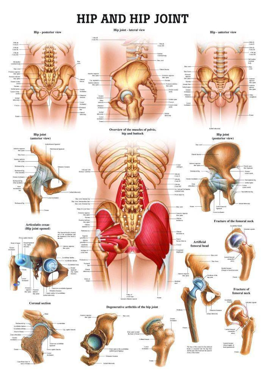
Hip And Hip Joint Laminated Anatomy Chart from cdn11.bigcommerce.com Causes of tightness a couple of the most obvious causes for muscle tightness in your hips and lower back are acute injuries — such as muscle strains — or simple soreness from doing more exercise than your body. Female hip muscles diagram ~ anatomy of the male and female pelvis comprehensive orthopaedics. If you are starting to feel hip pain or stiffness, you'll want to know more about the bones and muscles that make up the hip's anatomy. The sciatic nerve is the most commonly recognized nerve in the hip and thigh. Back muscle diagrams labeled 12 photos of the back muscle diagrams labeled back muscle diagrams labeled, lower back muscle diagrams labeled, human muscles, back muscle diagrams labeled, lower back muscle diagrams labeled. The four groups are the anterior group, the posterior group, adductor group, and finally the abductor group. These muscles can be grouped based upon their location and function. It is also referred to as a ball and socket joint and is surrounded by muscles, ligaments, and tendons.
The sacroiliac (si) joints connect the sacrum at the base of the spine with the hip bone.
ads/bitcoin2.txt
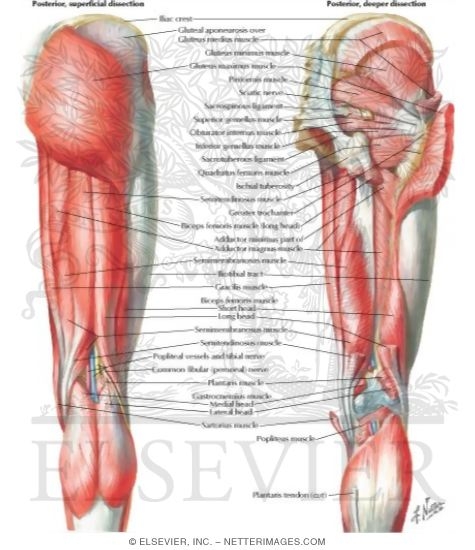
Ebraheim's educational animated video describes the muscle anatomy of the hip and buttocks region with simple images; Like the forearm, the upper leg, or thigh, has a dense arrangement of many muscles. Nerves carry signals from the brain to the muscles to move the hip and carry signals from the muscles back to the brain about pain, pressure and temperature. The iliacus and psoas major comprise the iliopsoas group. The sciatic nerve is the most commonly recognized nerve in the hip and thigh. Common causes of tight hip and lower back muscles include injury, too little activity, too much activity and muscular imbalances. Female hip muscles diagram ~ anatomy of the male and female pelvis comprehensive orthopaedics. Learn the iliopsoas, gluteal and hip adductors with diagrams now at kenhub In a nutshell, tight outer hip muscles, also known as. They provide a great deal of strength to modulate powerful forces between the upper and lower body. It is also referred to as a ball and socket joint and is surrounded by muscles, ligaments, and tendons. The thigh bone or femur and the pelvis join to form the hip joint. Back muscle diagrams labeled 12 photos of the back muscle diagrams labeled back muscle diagrams labeled, lower back muscle diagrams labeled, human muscles, back muscle diagrams labeled, lower back muscle diagrams labeled.
Five pairs of lumbar spinal nerves labeled l1 to l5 branch off your spinal cord and exit through small holes between the vertebrae. It runs from your lower back through your pelvis, passing to the front of your hip where it attaches to the top of your femur, which is your thigh bone. Common causes of tight hip and lower back muscles include injury, too little activity, too much activity and muscular imbalances. Nerves carry signals from the brain to the muscles to move the hip and carry signals from the muscles back to the brain about pain, pressure and temperature. On the anterior side, the most prominent of the muscles are the sartorius muscle and the four muscles that make up quadriceps muscle group (the quads.)
Muscles Of Back Of Hip And Thigh Muscles Of Hip And Thigh Posterior Views from netterimages.com Ebraheim's educational animated video describes the muscle anatomy of the hip and buttocks region with simple images; The muscles of the back are a group of strong, paired muscles that lie on the posterior aspect of the trunk they provide movements of the spine, stability to the trunk, as well as the coordination between the movements of the limbs and the back muscles are divided into two large groups: Together these muscles are commonly referred to as the iliopsoas. Muscles of the lower back and buttocks diagram 12 photos of the muscles of the lower back and buttocks diagram muscles of the lower back and buttocks diagram, human muscles, muscles of the lower back and buttocks diagram. The back muscles represented on an anatomical chart and on a schematic view of the origin and insertion of the proper muscles of the back (iliocostal muscle of the neck, lumbar (lumbar and thoracic parts), longissimus muscles of head, neck and thorax, the spinalis muscles of the neck and thorax, semispinalis muscle of the head, neck and thorax. Muscles of the lower back and buttocks diagram. The deep back muscles, also called intrinsic or true back muscles, consist of four layers of muscles: Superficial, intermediate, deep and deepest layers.these muscles lie on each side of the vertebral column, deep to the thoracolumbar fascia they span the entire length of the vertebral column, extending from the cranium to the pelvis
See back muscles and low back pain.
ads/bitcoin2.txt
To learn more about the anatomy of the spine, watch this video. Nerves in your lower back. These muscles can be grouped based upon their location and function. The part of the nerve that emerges out of the spine is called the nerve root. Like the forearm, the upper leg, or thigh, has a dense arrangement of many muscles. The bones together make up the hip. Learn the iliopsoas, gluteal and hip adductors with diagrams now at kenhub The psoas major is a large muscle that runs from the bodies and disc of the l1 to l5 vertebrae, joins with the iliacus via its tendon, and connects to the lesser trochanter of the femur. Muscles of the gluteal region. Diagram demonstrating the posterior view of the piriformis muscle orientation, origin and insertion on the pelvis and femur. The sacroiliac (si) joints connect the sacrum at the base of the spine with the hip bone. The psoas muscle is a deep muscle that connects your spine to your leg.in fact, it's the only muscle that does so. The hip abductors consist of the:
ads/bitcoin3.txt
ads/bitcoin4.txt
ads/bitcoin5.txt
0 Response to "Diagram Of Hip.and Back.muscles : Iliopsoas Wikipedia"

0 Response to "Diagram Of Hip.and Back.muscles : Iliopsoas Wikipedia"
Post a Comment