Leg Bones Diagram / Hip & Thigh - Atlas of Anatomy
Leg Bones Diagram / Hip & Thigh - Atlas of Anatomy. If you enjoyed learning the muscles of the leg with our quizzes and labeling exercises, look no further than our library of free quiz guides on tricky exam topics like the cranial nerves, bones of the skull and reproductive systems. Depending on the origin of the discomfort, upper leg pain symptoms can be a chronic nuisance or acute and debilitating. At the same time, the bones and joints of the leg and foot must be strong enough to support the body's weight while remaining. Look at links below to get more options for getting and using clip art. Also called the shin bone, the tibia is the longer of the two bones in the.
ads/bitcoin1.txt
The lower leg extends from the knee to the ankle. The muscles of the leg is a big topic, so take your time to learn it fully. It lies within the quadriceps tendon. Beside that, we also come with more related ideas as follows free printable human anatomy coloring pages, lower leg muscle diagram blank and lower limb bones unlabeled. The patella (kneecap) is the sesamoid bone in front of the knee.
LO_3943 Diagram Of The Lower Arm Bone Wiring Diagram from static-cdn.imageservice.cloud Bone diagram forehead (frontal bone) nose bones (nasals) cheek bone (zygoma) upper jaw (maxilla) lower jaw (mandible) breast bone (sternum) upper arm bone (humerus) lower arm bone (ulna) thigh bone (femur) collar bone (clavicle) toe bones (phalanges) ankle bones (tarsals) kneecap (patella) shin bone The back of the patella is covered with smooth cartilage. 10 october 2007 (original upload date) source: Human body anatomy human anatomy and physiology anatomy bones leg anatomy leg bones musculoskeletal system medical coding medical field sports medicine. (note, the radius and ulna bones also have this membrane.) this membrane keeps the tibia and fibula together and provides strength and stability for them. Blank leg bones diagram : With different grades of sprains depending on severity. The femur, or thighbone, is the longest and largest bone in the human body.
The back of the patella is covered with smooth cartilage.
ads/bitcoin2.txt
This area is commonly referred to as the calf. By tightening and relaxing, the skeletal muscles create movement. The major bones of the leg are the femur (thigh bone), tibia (shin bone), and adjacent fibula, and these are all long bones. With different grades of sprains depending on severity. The back muscles are skeletal muscles. The femur, or thighbone, is the longest and largest bone in the human body. This large tendon from the powerful thigh muscles (quadriceps) wraps round the patella and is attached to the top of the lower leg bone (tibia). Bone diagram forehead (frontal bone) nose bones (nasals) cheek bone (zygoma) upper jaw (maxilla) lower jaw (mandible) breast bone (sternum) upper arm bone (humerus) lower arm bone (ulna) thigh bone (femur) collar bone (clavicle) toe bones (phalanges) ankle bones (tarsals) kneecap (patella) shin bone It lies within the quadriceps tendon. Also called the shin bone, the tibia is the longer of the two bones in the. The knee joint is the largest joint in the body and is primarily a hinge joint, although some sliding and rotation occur. It also separates muscles on the anterior and posterior parts of the leg. Most of the leg skeleton has bony prominences and margins that can be palpated and some serve as anatomical landmarks that define the extent of the leg.
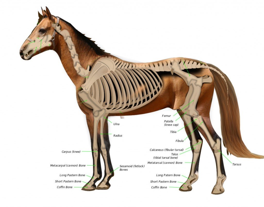
The bones of the leg are the femur, tibia, fibula and patella.the foot bones shown in this diagram are the talus, navicular, cuneiform, cuboid, metatarsals and calcaneus. Labeled human leg bones created for use in leg bone. Human body anatomy human anatomy and physiology anatomy bones leg anatomy leg bones musculoskeletal system medical coding medical field sports medicine. This large tendon from the powerful thigh muscles (quadriceps) wraps round the patella and is attached to the top of the lower leg bone (tibia). At the same time, the bones and joints of the leg and foot must be strong enough to support the body's weight while remaining.
Arm Bone Diagram . Arm Bone Diagram Upper Leg Bone Diagram ... from i.pinimg.com This image is an edited version of this image that was created by user:ladyofhats (mariana ruiz villarreal). The tibia and fibula are two long bones that run parallel to each other, forming the scaffold of the leg and providing attachment points for many muscles. Bone diagram forehead (frontal bone) nose bones (nasals) cheek bone (zygoma) upper jaw (maxilla) lower jaw (mandible) breast bone (sternum) upper arm bone (humerus) lower arm bone (ulna) thigh bone (femur) collar bone (clavicle) toe bones (phalanges) ankle bones (tarsals) kneecap (patella) shin bone There are two types of cartilaginous joints: There are in all 7 bones, which fall under tarsal bones category. It lies within the quadriceps tendon. The quadriceps muscle attachment points. With different grades of sprains depending on severity.
The bones of the leg are the femur, tibia, fibula and patella.the foot bones shown in this diagram are the talus, navicular, cuneiform, cuboid, metatarsals and calcaneus.
ads/bitcoin2.txt
Related posts of muscles and tendons of the leg shoulder muscle anatomy image. Foot and leg bones diagram best secret wiring diagram. The tibia and the fibula, at the top of the ankle joint. (note, the radius and ulna bones also have this membrane.) this membrane keeps the tibia and fibula together and provides strength and stability for them. It also separates muscles on the anterior and posterior parts of the leg. If you enjoyed learning the muscles of the leg with our quizzes and labeling exercises, look no further than our library of free quiz guides on tricky exam topics like the cranial nerves, bones of the skull and reproductive systems. The bones of the leg and foot form part of the appendicular skeleton that supports the many muscles of the lower limbs. Beside that, we also come with more related ideas as follows free printable human anatomy coloring pages, lower leg muscle diagram blank and lower limb bones unlabeled. The back of the patella is covered with smooth cartilage. I would think they would do one at s time but do not have. Now let's look at the tibia bone, which is the larger of the two leg bones, located medially. The major bones of the leg are the femur (thigh bone), tibia (shin bone), and adjacent fibula, and these are all long bones. The foot bones shown in this diagram are the talus, navicular, cuneiform, cuboid, metatarsals and calcaneus.
The knee joint is the largest joint in the body and is primarily a hinge joint, although some sliding and rotation occur. The tarsal bones in the foot are located amongst tibia, metatarsal bones, and fibula. Also called the shin bone, the tibia is the longer of the two bones in the. Foot and leg bones diagram best secret wiring diagram. Labeled human leg bones created for use in leg bone.
Why Do They Euthanize A Horse With A Broken Leg? » Science ABC from www.scienceabc.com Labeled human leg bones created for use in leg bone. Its lower end helps create the knee joint. Shoulder muscle anatomy image 12 photos of the shoulder muscle anatomy image shoulder muscle anatomy images, shoulder muscle anatomy picture, human muscles, shoulder muscle anatomy images, shoulder muscle anatomy picture The rounded, proximal end is the head of the femur, which articulates with the acetabulum of the hip bone to form the hip joint. Original file at image/png format. Every skeletal muscle has three main parts: Most of the leg skeleton has bony prominences and margins that can be palpated and some serve as anatomical landmarks that define the extent of the leg. Human body anatomy human anatomy and physiology anatomy bones leg anatomy leg bones musculoskeletal system medical coding medical field sports medicine.
The knee joint is the largest joint in the body and is primarily a hinge joint, although some sliding and rotation occur.
ads/bitcoin2.txt
The rounded, proximal end is the head of the femur, which articulates with the acetabulum of the hip bone to form the hip joint. With different grades of sprains depending on severity. The major bones of the leg are the femur (thigh bone), tibia (shin bone), and adjacent fibula, and these are all long bones. Its lower end helps create the knee joint. The tibia and the fibula, at the top of the ankle joint. These muscles work together to produce movements such as standing, walking, running, and jumping. The patella is the kneecap bone. There are in all 7 bones, which fall under tarsal bones category. The knee joint is the largest joint in the body and is primarily a hinge joint, although some sliding and rotation occur. Human body anatomy human anatomy and physiology anatomy bones leg anatomy leg bones musculoskeletal system medical coding medical field sports medicine. The patella (kneecap) is the sesamoid bone in front of the knee. Also called the shin bone, the tibia is the longer of the two bones in the. The femur, or thigh bone, is the single bone of the thigh region (figure 6.51).
ads/bitcoin3.txt
ads/bitcoin4.txt
ads/bitcoin5.txt
0 Response to "Leg Bones Diagram / Hip & Thigh - Atlas of Anatomy"


0 Response to "Leg Bones Diagram / Hip & Thigh - Atlas of Anatomy"
Post a Comment